Human Eye

*Eyes help to see the objects
*Eyes are situated inside a body cavity of the skull called
Ans : Orbits
*Study of eye and eye diseases
Ans : Ophthalmology
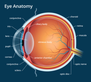
*There are three layers present in the eye ball
Ans : Sclera, Choroid, Retina
*The transparent front portion of sclera is known as
Ans : Cornea
*The middle layer of eye, nourishes oxygen and food
Ans : Choroid
*Behind the cornea the front portion of choroid, hangs like a vertical curtain called
Ans : Iris
*The opening seen at the centre of iris is called
Ans : Pupil
*The convex lens is present just behind the
Ans : Pupil
*The innermost layer of eye where the image is formed
Ans : Retina
*The space between lens and cornea is called
Ans : Aqueous chamber
*Aqueous chamber is filled with
Ans : Aqueous humour
*Aqueous humour supplies oxygen and nutrition for
Ans : Lens and cornea
*The space between lens and retina is called
Ans : Vitreous chamber
*Vitreous chamber is filled with
Vitreous humour
*Vitreous humour helps to maintain the shape of
Ans : Eyeball
*The ‘safe guards of eye’
Ans : Eyelids
*Yellow spot (fovea) is seen in
Ans : Retina
*The area of keenest vision and the region is characterised by the presence of cones only
Ans : Yellow spot
*Outer layer of eye – Sclera
* Middle layer of eye – Choroid
*Inner layer of eye – Retina
*The cells responsible for dim light vision
Ans : Rods cells
*The pigment present in rod cells
Ans : Rhodopsin
*Rhodopsin is called
Ans : Visual purple
*The compound obtained from vitamin A help to synthesize Rhodopsin
Ans : Retinin
*The poor vision in Dim light is caused due to the deficiency of
Ans : Vitamin A
*The poor vision in Dim light is known as
Ans : Night Blindness
*Cone cells help to percept the colours and cone cells contain a pigment called
Ans : Photospin
*Cells responsible for bright light vision and colour vision
Ans : Cone Cells
*The enzyme present in tears are
Ans : Lysozymes
*If the distant object is looked at fixedly, a clear image is formed in
Ans : Yellow spot
*The right distance which enable the proper vision is
Ans : 25cm
*The metal responsible for brightness of eye
Ans : Zinc
*The metal seen in tear is
Ans : Zinc
*The lens present in eye is
Ans : Biconvex lens
*The lachrymal glands produce
Ans : Tears
EYE DISORDER

*The disease caused by a reduction in the elasticity of lens, with age is called
Ans : Presbyopia
*The lens becomes either partially or completely opaque with age
Ans : Cataract
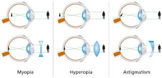
*The condition of not seeing distant objects clearly since the image is formed in front of the retina
Ans : Short-sight
*Short- sight is otherwise known as
Ans : Myopia (Near- sightedness)
*The defect of short- sight is corrected by using
Ans : Bi concave lens
*The condition of not seeing near objects clearly since the image is formed behind the retina
Ans : Long-Sight
*Long-sight is otherwise known as
Ans : Hypermetropia
*The defect of long- sight is corrected by using
Ans : Bi convex lens
*The condition of curvature of cornea become irregular and the image is not clearly formed
Ans : Astigmatism
*The defect of Astigmatism is corrected by using
Ans : Cylindrical lens
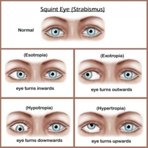
*The condition in which the eyes do not properly align with each other when looking at an object
Ans : Crossed eye / Strabismus /Squint Eye
*Squint eye is otherwise known as
Ans : Crossed eye / Strabismus
*The defect of squint eye is corrected by
Ans : Eye surgery
*The condition due to the increase of j pressure in the eye ball
Ans : Glaucoma
*Pain in eyes and seeing halos around light are due to
Ans : Glaucoma
*Inflammation of the outermost layer of the white part of the eye and the inner surface of the eyelid
Ans : Conjunctivitis (pink eye)
*Disable to distinguish the colours is known as
Ans : Colour Blindness
*A person who suffers colour blindness cannot distinguish
Ans : Red and Green
*Colour blindess is also known as
Ans : Daltonism
*Colour blindness was discovered by
Ans : John Dalton
*The procedure of replacing abnormal corneal tissue with a healthy cornea is known as
Ans : Keratoplasty
*The newly discovered layer in human cornea is
Ans : Dua’s layer
*Dua’s layer is discovered by the Indian Scientist
Ans : Harminder Singh Dua
*The abnormal protrusion of the eyeball or eyeballs is called
Ans : Exophthalmos
*The visual activity can be measured using an eye chart called
Ans : Snellen chart
The retention of a visual image for a second after the removal of the object is called
Persistence of vision The instrument used to examine the inner eye
Ophthalmoscope
The first eye transplant surgery was done by
Edward Konard Sim (1905)

“My Notebook ” is providing e books for biology lessons. Rs 20/- only. if you want to get it you can contact me in the form below.
- [PDF] Syllabus Law Officer Kerala State Co-operative Societies|192/2025 syllabus Kerala PSC
- [PDF] Syllabus Assistant Manager Kerala Welfare Fund Board KWFB| 433/2024 syllabus Kerala PSC
- Padma Awards 2026|Padma Vibhushan Awards 2026|Padma Bhushan Awards 2026|Padma Shri Awards 2026
- [PDF]Syllabus Assistant Director National Savings Kerala PSC|698/2025 Syllabus Kerala PSC
- [PDF] Syllabus HST Hindi|134/2025 syllabus Kerala PSC

